The 19 Muscles Of The Foot - PODIATRIST DESCRIBES HOW LEG AND FOOT MUSCLES INTERACT / Muscles of the ankle and foot.. (a) the insertions of the flexor digitorum longus, flexor hallucis longus and little attention has been paid to the clinical assessment of intrinsic foot muscles in the musculoskeletal injury literature apart from few specific. It arises from the femur; (from schuenke m, schulte e fig. Descriptive anatomy divides the skeletal elements of the foot into the tarsus, metatarsus, and forefoot (antetarsus). 10.19 (a) pattern of peripheral sensory innervation in the right lower limb.
A generous moment arm of these muscles about the midfoot. Several of the pims span the la in parallel with the plantar. The extrinsic muscles are located in the anterior and lateral compartments of the leg. There is a printable worksheet available for download here so you can take the quiz with pen and paper. Although prosthetics have come a long way, the complexity of the human foot and ankle mean it is hard to replicate.
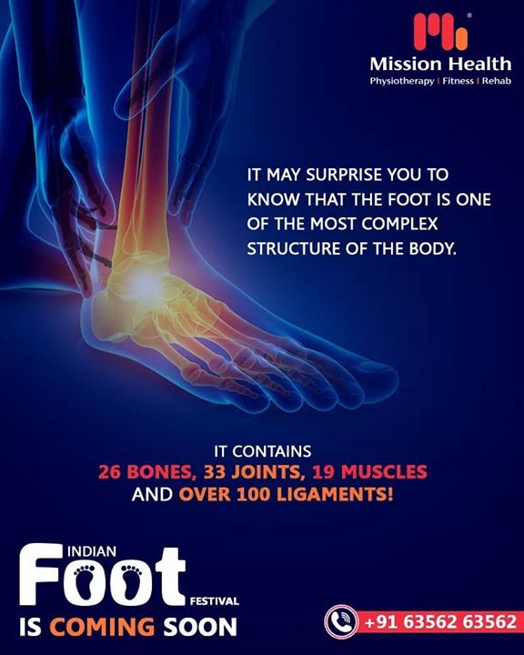
Anatomy Kickball by Jymeelah Kirk from img.haikudeck.com This article outlines the basic anatomy of the foot bones. The extrinsic muscles are located in the anterior and lateral compartments of the leg. The foot is an intricate part of the body, consisting of 26 bones, 33 joints, 107 ligaments, and 19 muscles. The dorsal aponeurosis of the toes supports the effect of the dorsal foot muscles by redirecting the force line of their tendons to. Their limited impact on posture and movement has led to the broad use of the extensor hallucis brevis and extensor digitorum brevis as muscular sources for tissue grafts. Flexion of 4 lesser toes at metatarsophalangeal, proximal & distal interphalangeal joints inversion of foot plantar flexion of ankle. Medial and lateral processes of posterior calcaneal tuberosity. To get started, all you need to do is click on the title of the article below that you are most interested in.
They are considered voluntary muscles.
This is an online quiz called muscles of the foot. Although prosthetics have come a long way, the complexity of the human foot and ankle mean it is hard to replicate. The muscles acting on the foot can be divided into two distinct groups; The bones and joints in the feet experience wear and tear, so conditions that cause damage to the foot can directly affect its health. Functional and clinical criteria divide the pedal skeleton into hindfoot, midfoot, and forefoot. Some run together to form complex webs around areas which need extra support, such as the sole of the foot, the top of the. Explore the muscles of the foot in this complete guide! They retract the foot and effect. There is a printable worksheet available for download here so you can take the quiz with pen and paper. They are generally divided into two sets: As a result, during walking the body's center of gravity normally fluctuates only 5cm in both vertical and lateral directions. Sides of adjacent metatarsals i: Gastrocnemius is a muscle of the posterior leg, where it forms a large part of the calf.
The muscles at the top of the foot fan out to supply the individual toes. They are considered voluntary muscles. All content on and from osmosis is intended for educational and informational purposes only. The muscles acting on the foot can be divided into two distinct groups; Muscles that move the ankle and foot muscles that move the ankle and foot are shown in figures 7.19 and 7.20.
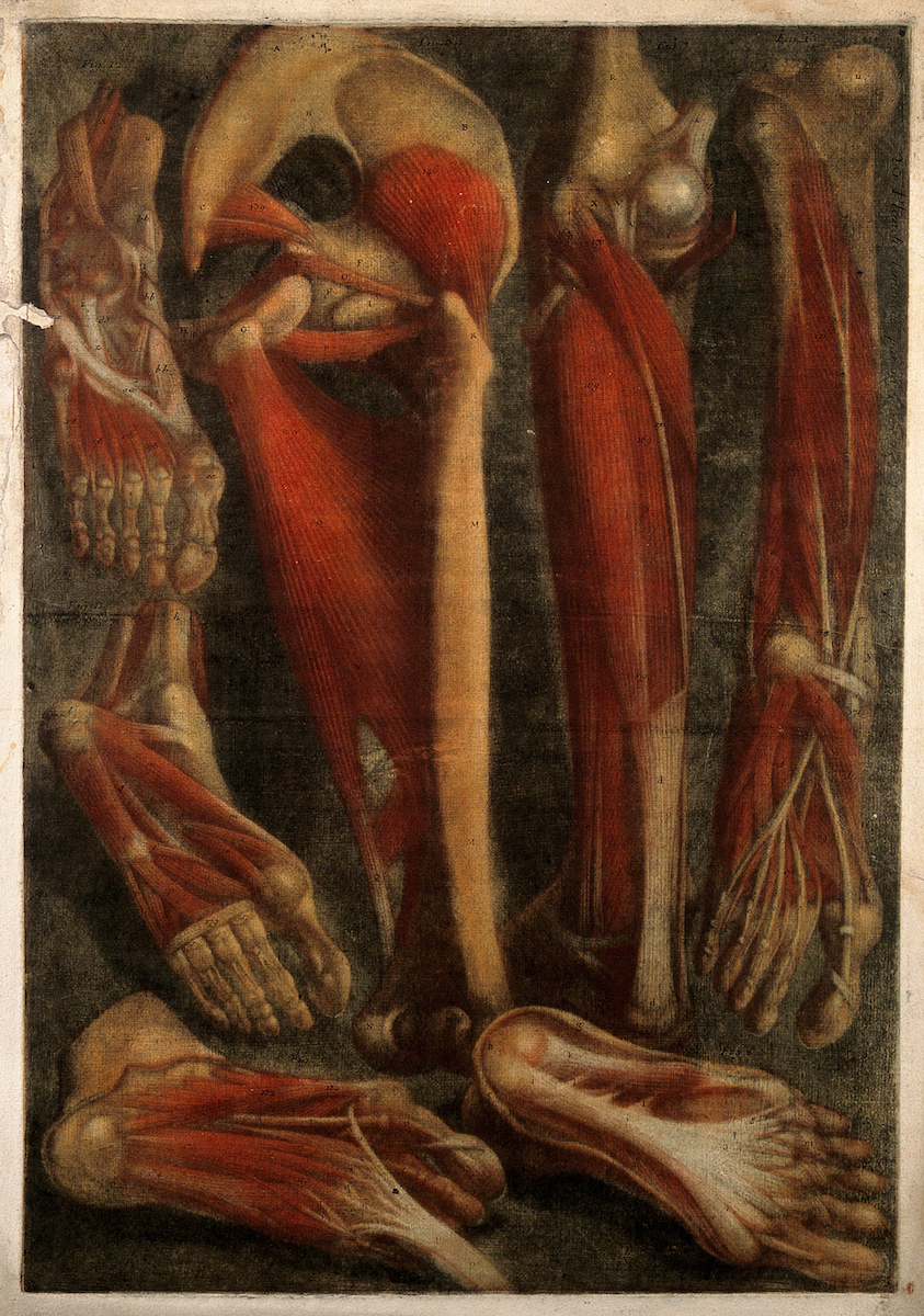
Muscles of the Foot and Leg - Product - The Public Domain ... from the-public-domain-review.imgix.net Distally, the muscle joins the strong calcaneal tendon, which attaches to the. Quiz which has been attempted 777 times by avid quiz takers. 4 in each foot, each with 2 heads o: This means that the little toe can only be extended by the extensor digitorum longus muscle only. Interestingly the dorsal foot muscles generally have no insertion at the little toe. Sides of adjacent metatarsals i: The muscles at the top of the foot fan out to supply the individual toes. Those of the medial plantar region are connected with the great toe, and corrrespond with those of the thumb;
The muscles acting on the foot can be divided into two distinct groups;
The bones and joints in the feet experience wear and tear, so conditions that cause damage to the foot can directly affect its health. The muscles acting on the foot can be divided into two distinct groups; They are generally divided into two sets: Maximum isometric force for the main pims is 375 n. Descriptive anatomy divides the skeletal elements of the foot into the tarsus, metatarsus, and forefoot (antetarsus). The muscles at the top of the foot fan out to supply the individual toes. Explore the muscles of the foot in this complete guide! They retract the foot and effect. This article outlines the basic anatomy of the foot bones. 10.19 (a) pattern of peripheral sensory innervation in the right lower limb. This is an online quiz called muscles of the foot. Although prosthetics have come a long way, the complexity of the human foot and ankle mean it is hard to replicate. The extensor digitorum brevis muscle (sometimes edb) is a muscle on the upper surface of the foot that helps extend digits 2 through 4.
They retract the foot and effect. There are 26 bones in each foot, as well as 33 joints, 19 muscles, 10 8. The bivalve foot, unlike that of gastropods, does not have a flat creeping sole but is bladelike (laterally the muscles mainly responsible for movement of the foot are the anterior and posterior pedal retractors. Maximum isometric force for the main pims is 375 n. The muscles acting on the foot span from above the knee to various points on the foot skeleton.
Dr. Alap Shah Foot is 1 of the most complex structure of ... from draalapshah.com Muscles that move the ankle and foot muscles that move the ankle and foot are shown in figures 7.19 and 7.20. They are generally divided into two sets: There are many ligaments in the foot. Some run together to form complex webs around areas which need extra support, such as the sole of the foot, the top of the. Blood flow restriction therapy learn how to improve. Their limited impact on posture and movement has led to the broad use of the extensor hallucis brevis and extensor digitorum brevis as muscular sources for tissue grafts. Layer 3 of the foot. Medial and lateral processes of posterior calcaneal tuberosity.
Flexion of 4 lesser toes at metatarsophalangeal, proximal & distal interphalangeal joints inversion of foot plantar flexion of ankle.
Usmle® is a joint program of the federation of state medical boards. They retract the foot and effect. Their limited impact on posture and movement has led to the broad use of the extensor hallucis brevis and extensor digitorum brevis as muscular sources for tissue grafts. Gastrocnemius is a muscle of the posterior leg, where it forms a large part of the calf. 10.19 (a) pattern of peripheral sensory innervation in the right lower limb. The bones and joints in the feet experience wear and tear, so conditions that cause damage to the foot can directly affect its health. It arises from the femur; They are considered voluntary muscles. Descriptive anatomy divides the skeletal elements of the foot into the tarsus, metatarsus, and forefoot (antetarsus). Medial and lateral processes of posterior calcaneal tuberosity. Insertions of the extrinsic foot muscle tendons on the plantar surface of the foot. Some run together to form complex webs around areas which need extra support, such as the sole of the foot, the top of the. To get started, all you need to do is click on the title of the article below that you are most interested in.